
Download Printable Version
Lab 11: Organization of the Skeleton
PURPOSE:To review the organization of the skeleton, the major bones of the skeleton, and the terms used to describe the skeletal structures
LEARNING OBJECTIVES:
1. Distinguish between the axial and appendicular skeleton
2. Locate and name the major bones of the skeleton
3. Define the terms used to describe skeletal structures
PROCEDURE:
1. Review Cpt 6 pages 137-139, Table 6-1, and the Skeleton Outline page
2. Print out image 11.1 (Below), paste into your NoteBook and Label
3. Examine the human skeleton and locate the following parts. Palpate the corresponding bones in your own skeleton, if possible: AXIAL SKELETON: skull: cranial bones, facial bones; hyoid; vertebral column: vertebrae, intervertebral disks, sacrum, coccyx; Thoracic cage: ribs, sternum; APPENDICULAR SKELETON: Pectoral Girdle: Scapula, Clavicle; Upper Limbs: Humerus, Radius, Ulna, Carpals, Metacarpals, Phalanges; Pelvic Girlde: Coxal Bones; Lower Limbs: Femur, Tibia, Fibula, Patella, Tarsals, Metatarsals, Phalanges. Use colored pencils to color the illustrations.
4. Locate an example of each of the following structures in the human skeleton. List them, the example and a brief definition in your NoteBook: Condyle, Crest, Epicondyle, Facet, Fontanel, Foramen, Fossa, Head, Meatus, Process, Sinus, Spine, Suture, Trochanter, Tubercle, Tuberosity
5. Complete Part A, B & C of the Lab Handout
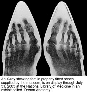
6. Doctors often use X-Rays to examine internal parts of the body to assist their diagnosis. Bone, in particular, shows up very well on film. Try you hand at reading X-Rays by examine the images of X-Rays below and try to ID as many bones as possible. Print out the images and paste them into your NoteBook, labeling them as you go.
7. As you complete the lab, Review the "Lab Objectives" from the handout and write a synopsis of the lab addressing the three objectives.
 |
 |
 |
 |
 |